Visual Structures: multiple specialized cortical areas concerned with different visual attributes such as “motion,” “color,” and other dimensions. (Ramachandran, 102) Now we can study the part of your visual brain that responds just to the visual stimulus. Experiments have shown that you can have actual brain imaging activity in the visual part of the brain (visual cortex) although you yourself are not conscious of the activity. It tells us that it's just not true that any activity in the (cerebral) cortex automatically becomes conscious. (Koch, BSP22)
When a person’s eyes are damaged, “signals” no longer flood in along the pathways to the occipital cortex. So that part of the cortex becomes no longer visual. Its’ territory is taken over by (competitive functional areas) of sensory information. As a result, when a blind person passes her fingertips over the raised dots of a Braille poem, her occipital cortex becomes active from mere touch. And it’s not only touch, but any sources of information. The blind subjects listen to sounds, their auditory cortex becomes active, and so does their occipital cortex. Age matters. In those born blind, (the) occipital cortex is completely taken over by other senses. If a person goes blind at an early age— say at five years old— the takeover is less comprehensive. (Eagleman, 37-38) (Considering other structures), the “amygdala” has projections to all levels of the visual cortex. So, it is… able to… to influence, visual processing at many levels. Being able to influence visual processing … determines what we can see out there. (The amygdala helps guide) the visual system in determining what gets to be seen. It's… picking out… information that’s significant, from what is not as significant, and helping guide vision and visual processing. (Pessoa, BSP106)
Blind Spot: a spot in the visual field in which vision is absent or deficient. (GHR) A small portion of an eye’s visual field (that can't pick up visual information) due to the "optic disk." The brain uses its mechanism of “filling-in” to complete images across the blind spot, however there is a limit to what can and cannot be filled in. (Ramachandran, 89) There are two eyes and blind spots are in different, non-overlapping locations; this means that with both eyes open you have full coverage of the scene. The brain fills in the missing information from the blind spot. (Eagleman, 33) Also referred to as “scotoma.”
Extrastriate Body Area (EBA): a brain region in the back of the brain, in the “parietal lobes” and “temporal lobes,” that responds to images of human bodies and body parts. Specializes in multi-sensory body-related information. Your EBA tracks other people’s EBA’s as well as your own. (Blakeslee, 204, 213)
Fovea: a small area in the center of the “retina,” composed entirely of “cones,” where visual information is most sharply focused. (Hockenbury, 91) Area consisting of a small depression in the retina containing only cones and where vision is most acute. (NCIt)
Fusiform Facial Area (FFA): brain area activated when we look at faces. (Hockenbury, 262)
Middle Temporal Area (MT): a small patch of “cortical” tissue found in each "hemisphere," that appears to be mainly concerned with seeing movement. (RamachandranTTB, 60)
Occulomotor Nerve Nucleus: adjacent to the “auditory nucleus,” this structure sends commands to the eyes, instructing them to move. (Ramachandran, 38)
Optic Chiasm: point in the brain where the “optic nerve” fibers from each eye meet and partly cross over to the opposite side of the brain. (Hockenbury, 92) An X-shaped structure formed by the two optic nerves, which pass backwards from the eyeballs to meet in the midline beneath the brain. (OxfordMed) At the optic chiasm the fibers from the medial part of each retina cross to project to the other side of the brain, while the lateral retinal fibers continue on the same side. As a result each half of the brain receives information about the contralateral visual field from both eyes. (MeSH) Also referred to as ‘optic decussation.’
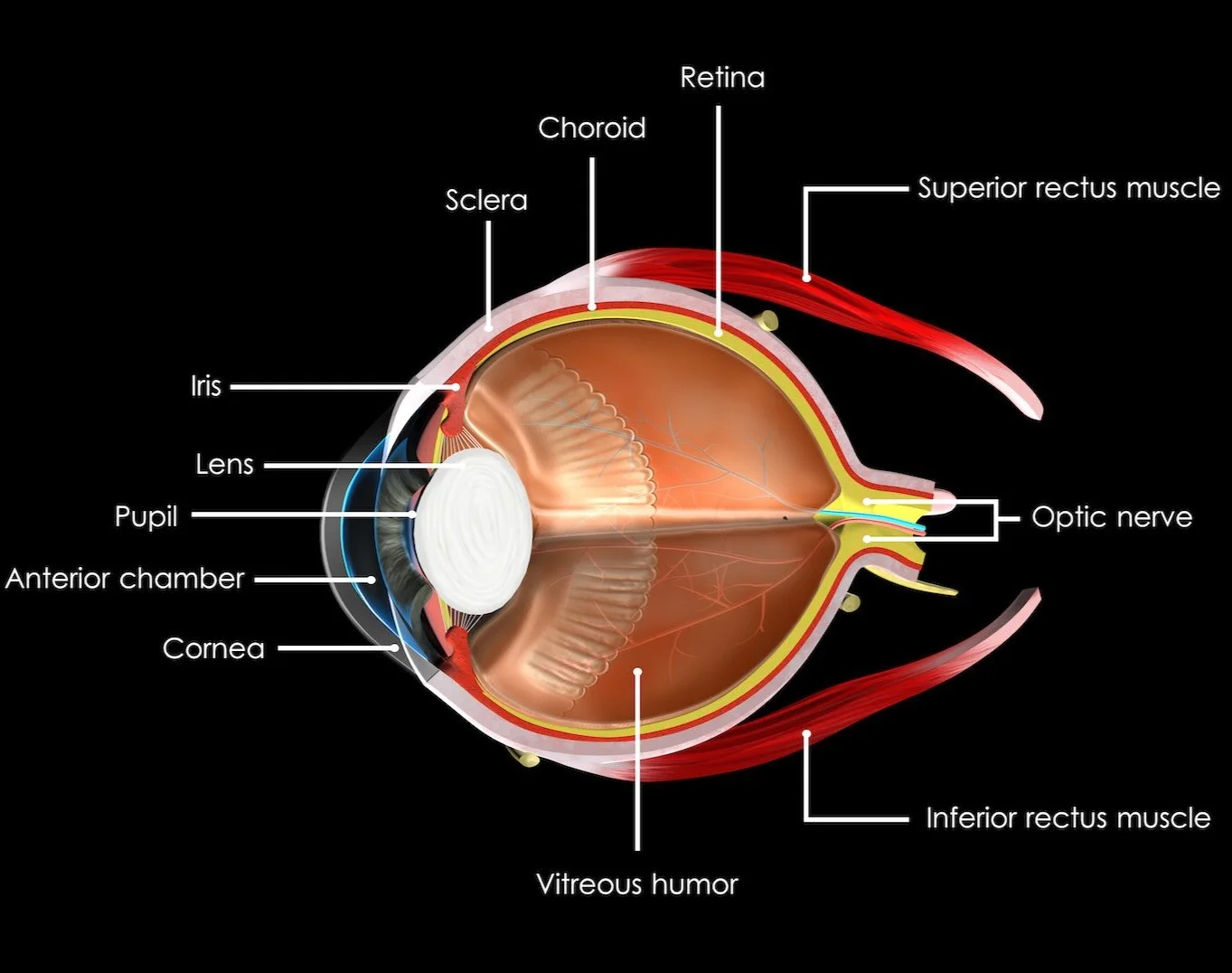
Optic Disk: the area of the retina where the optic nerve exits the eyeball. Unlike the other parts of the retina, it is not sensitive to light, so the result on vision is a blind spot. (Ramachandran, 89) A portion of the retina at which the axons of the "ganglion cells" exit the eyeball to form the optic nerve. No light-sensitive photoreceptors are contained within this portion of the retina. (NCIt) Area of the retina without “rods” or cones. (Hockenbury, 91)
Optic Nerve: the thick nerve that exits from the back of the eye and carries visual information to the visual cortex in the brain. (Hockenbury, 92) The 2nd cranial nerve. Conveys visual information from the retina to the brain. The nerve carries the axons of the retinal ganglion cells which sort at the optic chiasm and continue via the optic tracts to the brain. The largest projection is to the “lateral geniculate nuclei.” Other important targets include the “superior colliculi” and the ‘suprachiasmatic nuclei.’ Though known as the second cranial nerve, it is considered part of the “central nervous system.” (MeSH) The optic nerve comes in to the lateral geniculate nucleus (of the “thalamus”) and then sends its information to the “primary visual cortex.” (Bainbridge, BSP32)
Orbit: the cavity in the skull that contains the eye. (OxfordMed) Bony cavity that holds the eyeball and its associated tissues and appendages. (MeSH) Seven bones contribute to the structure of the orbit… (NCIt) Adjective - ‘orbital.’
Photoreceptors: two classes of these are spread unevenly across the “retina.” About one hundred million "rods" work best under dim light conditions, while five million "cones" mediate daylight vision. For most day-to-day activities (including reading), only the cones provide a reliable “signal.” (Koch, 51) When exposed to light, the rods and cones undergo a chemical reaction that results in a neural signal. (Hockenbury, 90)
Cones: one of the two photoreceptor cell types in the “vertebrate” retina. Cones are less sensitive to light than rods, but they provide vision with higher spatial and temporal acuity, and the combination of signals from cones with different pigments allows color vision. (MeSH) Short, thick, pointed sensory receptors of the eye that detect color and are responsible for color vision and "visual acuity." (Cones) require much more light than rods do, to function effectively. This is why it is difficult or impossible to distinguish colors in very dim light. Adapt quickly to bright light. Specialized for seeing fine details and for vision in bright light. (Hockenbury, 90-91)
Rods: one of the two photoreceptor cell types of the vertebrate retina. In rods the photopigment is in stacks of membranous disks separate from the outer cell membrane. (MeSH) Long, thin, blunt sensory receptors of the eye that are highly sensitive to light, but not to color, and that are primarily responsible for ‘peripheral vision’ and ‘night vision.’ Once the rods are fully adapted to the dark, they are about a thousand times better than cones at detecting weak visual stimuli. We therefore rely primarily on rods for our vision in dim light and at night. Rods adapt relatively slowly to changes in the amount of light. (Hockenbury, 90-91)
Primary Visual Cortex (V1): located in the “occipital lobes” along the banks of a deep “sulcus” called the “calcarine fissure.” (Blumenfeld, 28) The first and largest of our cortical visual maps. (RamachandranTTB, 63) Involved in visual inputs. (Hawkins, 113) This bit of cortex is a region of multiple sensory inputs and multiple outputs. In addition, each single input or output involves large numbers of individual “axons” over the entire area. The main function is to respond to the orientation of “forms” in the visual field. (The Brain-Francis Crick, 135) Takes in images. (Goldberg, 30) Like a war room where information is constantly being sent back from scouts. (Ramachandran, 110) (In addition to) the visual cortex, more than 30 areas in the brains of primates - including humans - are involved in handling aspects of vision such as the perception of motion, color, and depth. (SAM Dec08/Jan09, 20) Input that arrives (into V1) is segregated into three separate types of information: color, form, and movement. This information is then sent to V2. (Kolb, 283) When damaged, cortical blindness develops even though the eyes continue to work fine. (Goldberg, 24) Also referred to as ‘visual cortex,’ ‘occipital cortex,’ ‘striate cortex,’ 'Brodmann area 17,' and ‘V1.’
Retina: a thin, light-sensitive membrane located at the back of the eye that contains the “receptor cells” for vision. (Hockenbury, 91) The ten-layered nervous tissue membrane of the eye. (MeSH) The light-sensitive layers of nerve tissue at the back of the eye that receive images and send them as electric signals through the optic nerve to the brain. (NCIt)
Secondary Visual Cortex: a group of cortical areas surrounding the visual cortex. (Koch, 335) After the primary visual cortex, the remaining areas of the “occipital lobe.” Because each of these occipital regions has a unique cellular structure and has unique inputs and outputs, we can infer that each must be doing something different from the others. (Kolb, 282) Also referred to as ‘V2,’ ‘V3,’ ‘V4,’ ‘V5,’ and ‘extrastriate cortex.’
V2: one of the thirty visual areas in the brain. (RamachandranTTB, 61) Here, color, form, and movement inputs (received from V1) remain segregated. Visual pathways proceed from region V2 to the other occipital regions and then to the parietal and temporal lobes. (Kolb, 283)
V4: an area in the temporal lobes that appears to be specialized for processing color. When this area is damaged on both sides of the brain, the entire world looks like a black-and-white motion picture. But the patient’s other visual functions seem to remain perfectly intact. (RamachandranTTB, 61) This is different from the more common form of congenital “color blindness” that is due to eye problems. (Ramachandran, 73)
