Neuron Structures: the architecture of the neuron helps us begin to understand why the brain is so multifunctional, while organs like the "pancreas" and "spleen" are not. (LeDoux, 40) The nervous system contains neurons in an array of shapes and sizes, structured differently because of their specialized tasks. (Kolb, 79)
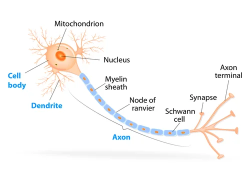
Each neuron has three main elements. The “cell body,” which is the cell’s powerhouse and includes the cell “nucleus” and “organelles.” The main output fiber, known as the “axon,” arises from the cell body. Input fibers, known as “dendrites” stick out from the cell body a bit like antlers. (Damasio, 302)
Axon: one of the three main (components) of the neuron. A projection from the neuron "cell body” that can reach long distances in the brain. The axon’s function is to send data out of the cell. (Ramachandran, 9) Axons are "output" channels and dendrites are "input" channels. (LeDoux, 40) A protrusion emanating from the cell body that along with another neuron’s “dendrite” makes possible the connections between neurons. (Goldberg, 39) Long fiber of the neuron, often with thousands of contact points. (The Brain-Francis Crick, 132) Emerges at one end of the cell body and can extend up to several feet. Often splits into one or more branches along its length. (Kandel, 64) Most neurons have only axon. However, each axon branches many times before it ends, allowing a single neuron to spawn many terminals. The result is that messages sent out from one cell can affect many others. (LeDoux, 42) A critical part of the neuron. Extends away from the body of the neuron. Provides the pathway over which signals can travel from the cell body, sometimes over long distances to neurons in other parts of the brain, and in the nervous system. (The Brain-Charles Stevens, 15) A living cable of varying lengths (from microscopic up to six feet long). Often compared to wires because they carry electrical impulses at very high speeds (from 2 to 200 miles per hour) toward the dendrites of neighboring neurons. (Doidge, 53) Also referred to as ‘fiber,’ ‘nerve fiber,’ ‘process,’ and ‘cell process.’
Axon Collaterals: branches that diverge from the main axon at right angles. (Patestas, 30) Also referred to as ‘collaterals.’
Axon Hillock: juncture of the (neuron cell body) and axon. Where the action potential begins. (Kolb, 79)
Axon Membrane: the membrane that surrounds the axon. Contains special openings, known as “ion channels,” that allow "potassium" “ions” to flow from the inside of the cell, where they are present in high concentrations, to the outside, where they are present in low concentrations. Since potassium is a positively charged ion, its movement out of the cell leaves the inside surface of the membrane with a slight excess of negative charge. The outside surface of the cell membrane becomes lined with positive charges from the potassium ions that have diffused out of the cell. The inside of the membrane becomes lined with negative charges from “proteins” (inside the cell) attempting to draw potassium ions back into the cell. This balance of ions maintains the stable “resting membrane potential” of 70 millivolts. (Kandel, 80)
Axon Terminal: tiny end of each branch of an axon. (Kandel, 64) Enables communication with other neurons. (Ramachandran, 9) Contains neurotransmitters in synaptic vesicles. (NCIt) The point at which the sending neuron communicates with receiving neurons. (LeDoux, 40) At its terminus the axon may “arborize,” forming numerous axon terminals which permits a single axon to make synaptic contact with numerous other neurons, muscle cells or gland cells. (Patestas, 30) Also referred to as ‘terminal,’ ‘nerve terminal,’ ‘synaptic terminal,’ ‘presynaptic terminal,’ ‘presynaptic knob,’ ‘end foot,’ ‘terminal bouton,’ and ‘bouton terminaux.’
Presynaptic Membrane: membrane on the transmitter-output side of a synapse. Forms the axon terminal. (Kolb, 153)
Synaptic Vesicles: small secretory vesicles that contain a neurotransmitter, are found inside an axon near the presynaptic membrane, and release their contents into the “synaptic cleft” after fusing with the membrane. (GHR) Contain from 10,000 to 100,000 molecules of a specific type of neurotransmitter. There may be thousands of synaptic vesicles in a single terminal. The vesicles serve to protect the transmitter molecules from “enzymes” inside the terminal that would otherwise destroy them. (The Brain-Leslie Iversen, 76) Contain neurotransmitters. At the synapse, an electrical impulse triggers the "migration" of vesicles. The membrane of the vesicle fuses with the membrane of the terminal. This action releases neurotransmitters into the synaptic (cleft). (Chudler, 15) Characterized by a single “phospholipid” membrane. They transport substances through, into, and out of the cell. Produced by the “endoplasmic reticulum,” the “golgi apparatus,” and the cell membrane. Vesicles from the transmitting cell fuse to its membrane, releasing regulator chemicals by “exocytosis” into the “extracellular fluid." (Norton Lectures, 6/2/09) So finely miniaturized is the cellular structure of the nervous system that the length of a wave of visible light is too blunt to probe it. The wavelength of green light is ten times longer than the size of a synaptic vesicle. (Fields, 17) Also referred to as "vesicles" and “transport vesicles.”
Cell Body: the enlarged portion of a neuron containing the “nucleus.” (OxfordMed) Involved in important housekeeping functions such as storing genetic material and making proteins and other molecules that are necessary for the cell’s survival. (LeDoux, 40) The most prominent feature is the nucleus which possesses a fine “chromatin” network and a well-defined “nucleolus.” The “cytoplasm” is rich in free “ribosomes” and “rough endoplasmic reticulum.” Protein synthesis occurs on ribosomes for use in the “cytosol” and on the rough endoplasmic reticulum for eventual packaging. The “Golgi complex” is responsible for the modification and packaging of the various proteins, enzymes, and chemical messenger molecules manufactured on the rough endoplasmic reticulum. The energy requirement of the neuron is met by the presence of numerous “mitochondria,” which are distributed throughout the cell body. As such, “protein synthesis,” “respiration,” and many of the essential cellular functions occur in this region. (Patestas, 29) Material produced (here) is transported, via the assistance of “microtubules” to the axon for its use. Material may (also) be conveyed in the opposite direction, toward the neuron cell body. (Patestas, 30-31) Also referred to as a ‘soma,‘ ‘nerve body,’ ‘neuron body,’ and ‘neuronal body.’
Dendrites: resemble dense, twiggy thickets. (RamachandranTTB, 14) Branch extensively, forming a treelike structure that grows out from the cell body and spreads over a large area. Usually emerges on the opposite side of the cell body from the axon. (Kandel, 64) Often found intricately interlaced with other dendrites, even though they usually do not touch one another. (The Brain-Francis Crick, 132) The thinnest branches of neurons are less than a tenth of a “micron” in diameter, which is less than the wave length of visible light. (Cerebrum2009, 70) Receive input from other neurons. (Doidge, 52) Since dendrites are usually highly branched structures, they may receive information concurrently from many different sources. (Patestas, 30) There are three ways the (dendritic) tree influences how maw many spikes we need. The first is how far away from the neuron’s body the spike lands; the second is how bunched together on the tree are inputs from a chorus of neurons; the third is what lies ahead on the the path between the input and the body. (Humphries, 41) They also participate in “feedback loops.” (CampbellVA, 144) Also referred to as ‘dendritic tree.’
Dendritic Spine: little knobs extending from dendrites. Especially important as receivers of messages from axons. (LeDoux, 41-42) Delicate tube-like extension of the neuron. Dendrites and axons begin to develop during (pregnancy). Dendrites begin to sprout through the process called “arborization.” (Goldberg, 40)
Postsynaptic Membrane: membrane on the transmitter-input side of a synapse. (Kolb, 153) The membrane of the postsynaptic cell that is enriched with neurotransmitter “receptors.” (NCIt)