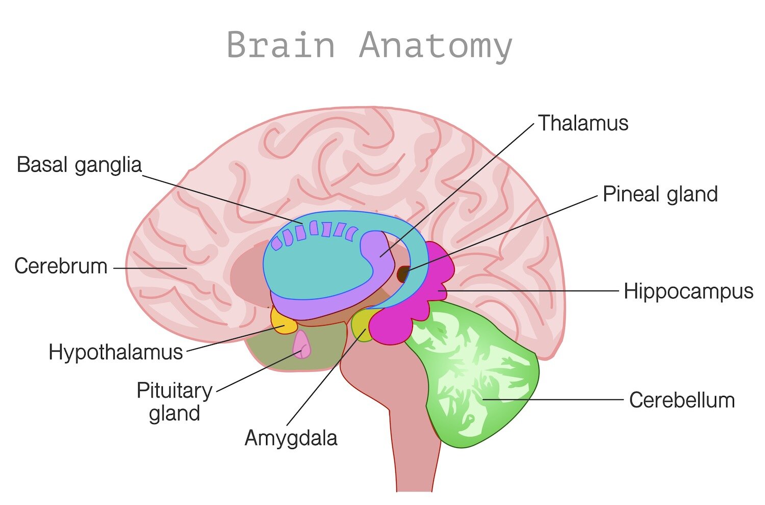
Memory Structures: ancient brain structures (much older than the cortex) involved in many crucial functions, including memory storage and spatial navigation. (Blakeslee, 213)
“Explicit memory” relies on the “medial” region of the “temporal lobe.” Rather than relying on higher, cognitive regions, “implicit memory” depends more on regions of the brain that respond to stimuli, for example the “amygdala,” the “cerebellum,” and the “basal ganglia.” (Kandel4, 111-112) Based on the study of “lesions” in humans and animals, two main regions of the brain appear to be critical to memory formation: the medial “temporal lobe” memory areas… and the medial “diencephalic areas.” The medial temporal lobe memory areas include the “hippocampal formation” and adjacent cortex of the “parahippocampal gyrus.” The medial “diencephalic” memory areas include the “dorsal-medial nucleus” (in the “thalamus”), the “anterior nuclear group” (in the thalamus), the “mammillary bodies” (in the “hypothalamus”), and other diencephalic “nuclei” lining the “third ventricle.” “White matter” network connections constitute a third component that is necessary for normal memory function. The “basal" forebrain may also play a role in memory. (Blumenfeld, 829)
Hippocampal Formation: one of several structures in the "limbic system." Buried within the medial temporal lobe, forming the floor of the temporal “horn” of the “lateral ventricle.” Extends from the “amygdala” to the “splenium” of the “corpus callosum.” Consists of “archicortex.” (Patestas, 346) Unlike the six-layered “neocortex,” it has only three layers. (Blumenfeld, 821) Involved in the consolidation of “short-term memory” into “long-term memory.” (Patestas, 344) Has an elaborate, curving ‘S’ shape on “coronal” sections. This appearance inspired the name “hippocampus” which means ‘sea horse’ in Greek. Three key structures include the hippocampus, the “dentate gyrus,” and the “subiculum.” (Blumenfeld, 830) (Has) many areas of input, including the parietal “association cortex,” the “occipital cortex,” and the “temporal cortex.” Outputs to the temporal and prefrontal (lobes) through the “Papez Circuit.” (Fisch, 378)
Dentate Gyrus: has a double-sided ‘C’-shape that cups the "superior" turn of the hippocampus. (Fisch, 374) Named for the toothlike bumps on its medial surface. Principal neurons are are called “granule cells.” (Blumenfeld, 830) A notched band of “cortex” that is interposed between the upper aspect of the “parahippocampal gyrus” and the “fimbria.” Consists of three layers of archicortex. 'Molecular layer' consists mainly of a small population of nerve cell bodies and granule cell "dendrites." 'Granule cell layer' contains the "cell bodies" of granule cells whose axons form the output of the dentate gyrus. 'Polymorphic layer' consists of “interneurons.” Neurons output to the hippocampus only. (Patestas, 349)
Hilus: an indentation on the surface of an organ where blood vessels, ducts, nerve fibers, etc., enter or leave it. (Oxford) Area of the dentate gyrus that consists of interneurons and granule cell axons. (Patestas, 349) Also referred to as 'hilum.'
Fornix: meaning ‘arch’ in Latin. White matter structure that curves through the “ventricular system” from the hippocampal formation to the “diencephalon” and “septal area.” Discrete bundle of output fibers... (Blumenfeld, 836) Heavily “myelinated” fiber bundle “projecting” from the hippocampal formation to the “hypothalamus.” Some authorities consider the fornix part of the limbic system. (MeSH) Also referred to as ‘fimbria.”
Hippocampus: memory forming part of the brain. “Grid” and “place cells” are linked here. Central to the “encoding” of memory. (Lynch, 77) Critical for learning new information. (Goldberg, 26) An infolding of the “cortex” of the human brain, embedded within the “parahippocampal gyrus” of the temporal lobe. (Patestas, 347) Involved in the complex processes of forming, sorting, and storing memories. (Chudler, 55) Resembles a ram’s horn. Named after the Egyptian god ‘Amon’ who bore a ram’s head. Sectioned into four regions ‘CA4 to CA1.’ CA2, CA3, and CA4 encompass large, ovoid-shaped neurons. The relative density of their (cell body) increases as you go from CA2 to CA4. (Fisch, 374) Includes two tiny seahorse-shaped structures. Controls the laying down of new memories. (Ramachandran, 15) The area that turns our memories from short-term to long-term ones. (Doidge, 97) Particularly vulnerable to “Alzheimer’s disease.” (Goldberg, 26) Imaging has found that, like many other disorders, “depression” may result in fewer and smaller synapses in the hippocampus. In fact, longer depressive episodes are correlated with reductions in the volume of the hipppocampus. This correlation would account for the problems with memory that people with depression experience. (Kandel4, 66) Editor’s note - named from the Greek word meaning ‘seahorse.' Plural - ‘hippocampi.’ Also referred to as ‘Ammon’s horn,’ and ‘cornu ammonis.’
CA1 Zone: located along the inferior portion of the superior turn of the hippocampus. Composed of triangular-shaped neurons. Is particularly susceptible to “anoxia.” (Fisch, 374) Lies next to the subiculum. (Patestas, 347) Also referred to as the ‘sommer sector.’
CA2 Zone: located along the descent of the superior turn of the hippocampus. (Fisch, 374)
CA3 Zone: located along the rise of the of the superior turn of the hippocampus. (Fisch, 374) Lies close to the dentate gyrus. (Patestas, 347)
CA4 Zone: positioned in the ‘concavity’ of the dentate gyrus. (Fisch, 374)
Pes Hippocampus: (the “anterior” extent of the hippocampus), has several groves resembling a paw. (Patestas, 347)
Subiculum: main output area (from the hippocampal formation) to the cerebral cortex. (Fisch, 378) Output (travels through) the hypothalamus, and “thalamus.” A transitional zone that displays a three-layered archicortex next to the hippocampus, but progressively becomes a more elaborate six-layered neocortex as it approaches the parahippocampal gyrus. Receives information relayed by the hippocampal pyramidal cells. (Patestas, 349) Also referred to as ‘subicular cortex.’
Parahippocampal Gyrus: lies above the “collateral sulcus.” (Fisch, 282) Key structure of the medial temporal lobe. (Blumenfeld, 831) Includes several cortical areas with connections to the hippocampal formation, the most important of which is the “entorhinal cortex.” (Blumenfeld, 833)
Entorhinal Cortex: the main input area from the cerebral cortex to the hippocampus. (Fisch, 378) Located on the medial surface of the temporal lobe. Provides a major route for neocortical input to the hippocampal formation. Often degenerates in Alzheimer’s disease. (Kolb, 497) “Stimulation” applied (here) while the subjects learned locations of landmarks, enhanced their subsequent memory of these locations: the subjects reached these landmarks more quickly and by shorter routes, as compared with locations learned without stimulation. (Suthana, Results)
Parahippocampal Cortex: cortex located along the dorsal medial surface of the temporal lobe. (Kolb, 497)
Parahippocampal Place Area (PPA): area of the brain activated when we look at pictures of places. (Hockenbury, 262)
Perirhinal Cortex: cortex lying next to the “rhinal (sulcus)” on the base of the brain. (Kolb, 497)
