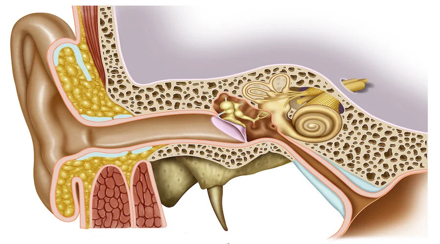
Auditory Structures: the organs involved with detecting and processing auditory information. (NCIt)
The ear collects "sound waves" from the surrounding air and converts them into electrochemical neural (signals) which then begin a long route through the "brainstem" to the "primary auditory cortex." (Kolb, 314) The ("cochlea") is the microphone inside our ears. When the external world produces sound, different "frequencies" vibrate different little hair cells within the cochlea. There are three thousand such hair cells, which convert the sound into “patterns” of electrical signals that travel down the "auditory nerve" to the (primary auditory cortex). (Doidge, 57)
Eustachian Tube: the tube that connects the middle ear to the "pharynx." It allows the pressure on the inner side of the eardrum to remain equal to the external pressure. (OxfordMed) Also allows middle ear secretions to drain into the pharynx. (NCIt)
Inner Ear: the essential part of the hearing organ. (MeSH) The part where sound is (converted) into impulses; consists of the “cochlea” and the “semicircular canals.” (Hockenbury, 96) Also referred to as ‘internal ear.’
Cochlea: the snail shell-shaped auditory component of the inner ear. It contains the sensory organ of hearing. (NCIt) A fluid-filled tube that's coiled in a spiral. (Hockenbury, 97) Concerned with the reception and analysis of sound. (OxfordMed) Adjective - ‘cochlear.’
Basilar Membrane: the membrane within the cochlea of the ear that contains the hair cells. The physical vibration of the sound waves is converted into neural impulses. (Hockenbury, 98-99)
Cochlear Hair Cells: embedded in the “basilar membrane,” they are the receptors for sound. Have tiny, projecting fibers that look like hairs. They bend as the basilar membrane ripples. As the hair cells bend, they stimulate the cells of the “auditory nerve,” which carries the neural information to the “thalamus” and the “auditory cortex” in the brain. (Hockenbury, 98)
Middle Ear: the part of the ear that consists of an air-filled space within the 'temporal' bone. It is lined with mucous membrane and is connected to the outer ear by the "eardrum." Within the middle ear are three bones which transmit sound vibrations from the "outer ear" to the inner ear. (OxfordMed) The eardrum’s vibration is transferred to these three tiny bones. Each bone sets the next bone in motion. The joint action of these three bones almost doubles the “amplification" of the sound. (Hockenbury, 97) Also referred to as 'tympanic cavity.'
Incus: a small anvil-shaped bone in the middle ear. (OxfordMed) One of the three bones comprising the middle ear. This anvil-shaped bone is positioned between the “malleus” and the “stapes.” (NCIt) Also referred to as 'anvil.'
Malleus: a hammer-shaped bone in the middle ear that (joins) with the incus and is attached to the eardrum. (OxfordMed) It is attached to the inner surface of the (ear drum) and its function is to transmit sound vibrations. (NCIt) Also referred to as 'hammer.'
Stapes: a stirrup-shaped bone in the middle ear that (joins) with the incus. (OxfordMed) Transmits sound vibrations from the incus to the internal ear. (MeSH) Also referred to as 'stirrup."
Outer Ear: the outer part of the hearing system of the body. (MeSH) The part of the ear that collects sound waves; consists of the “pinna,” the “ear canal,” and the “eardrum.” (Hockenbury, 96) Also referred to as ‘external ear.’
Ear Canal: the passage leading from the pinna of the outer ear to the "eardrum." (OxfordMed) Conducts the sound collected by the (pinna) to the (eardrum). (MeSH) Also referred to as 'auditory meatus.'
Eardrum: a tightly stretched membrane at the end of the ear canal that vibrates when hit by “sound waves.” (Hockenbury, 96) When sound waves reach the membrane, it vibrates transferring these vibrations to the middle ear to which it is attached. (OxfordMed) Also referred to as "tympanic membrane."
Pinna: a flap of skin and "cartilage" that projects from the head. In humans, the pinna may be partly concerned with detecting the direction of sound waves. (OxfordMed)
Oval Window: oval opening on the lateral wall adjacent to the middle ear. (MeSH) A membrane that separates the middle ear from the inner ear. Many times smaller than the eardrum. Receives the amplified vibration from the (stapes). As it vibrates, the vibration is next relayed to than inner structure called the cochlea. (Hockenbury, 97)
Primary Auditory Cortex: area of the temporal lobe concerned with hearing. (MeSH) The area in the cerebral cortex that receives and processes auditory input. (NCIt) Located deep in the “Sylvian fissure.” Arranged into two-dimensional, alternating, vertically oriented columns of neurons. (Patestas, 313) Responsible for processing auditory inputs. (Hawkins, 117) A brain region that helps recognize a sound for its source (for example waking to an alarm clock). It comes prewired with genetically transferred information. (Goldberg, 102) Sensory impulses generated in the inner ear first reach the primary auditory cortex via the “thalamus” and then enter the "secondary auditory cortex." Reacts specifically to the visual image of speech. Even the soundless image of a person speaking is sufficient to stimulate the auditory cortex, including when the speaker is talking gibberish. (SAM April/May07, 29) Imagining music can activate the auditory cortex almost as strongly as listening to it. Imagining music also stimulates the "motor cortex," and conversely, imaging the action of playing music stimulates the auditory cortex. (Sacks, 32) Also referred to as 'auditory cortex.'
Frequency Columns: cells in each “column” respond to an auditory stimulus of a particular “frequency.” (Patestas, 313)
Summation Columns: respond to an auditory stimulus that stimulates both ears simultaneously. (Patestas, 314)
Suppression Columns: respond maximally to an auditory stimulus that stimulates only one ear. (Patestas, 314)
Secondary Auditory Cortex: surrounds the primary auditory cortex. Region where (auditory) signals merge with other sensory information. The posterior end of the secondary auditory cortex is where sensory integration appears to occur. It appears that this end is specialized for registering “spatial” information – that is, recognizing the directionality of a sound. (SAM April/May07, 29) Plays an important function in the interpretation of sounds, and via connections with “Wernicke’s area,” functions in the comprehension of “language.” (Patestas, 314)
